Updated 1 week ago
Microscopic Specimen Observation Techniques | Microscopy Guide
sidrahafeez754

Microscopic Specimen Observation
A Comprehensive Guide to Microscopic Specimen Observation in Laboratory Science
Microscopic Specimen Observation is one of the most important techniques of laboratory science in the contemporary world. It helps scientists, technologists, and healthcare experts to get acquainted with the uncharted realm of cells, tissues, and microorganisms. This procedure contributes to scientific discovery, starting with the diagnosis of diseases, up to biological research.
The specimen is no longer a simple art in the laboratories to be viewed under the microscope. It is a methodical process that involves precision instruments, advanced imaging equipment, and careful planning strategies. At any level, whether it is microscopic observation of a biological specimen or microscopic examination of a specimen in the laboratory, precision is important.
Modern Approaches to Learning Microscopic Specimen Observation in Laboratories
Microscopic Specimen Observation refers to the observation of samples under optical or electronic microscopes in detail. It assists the scientists in studying the structures that are not visible to the naked eye.
This Includes:
• Cellular structures
• Tissue organization
• Microorganisms
• Disease-related changes
Examination of microscope specimens is a routine procedure in pathology, microbiology, and biomedical research. It helps in regular diagnostics and scientific research. Optical microscopy analysis and occasionally electron microscopy observation of biological samples give a high-resolution image of the biological sample in modern laboratories.
Importance of Microscopic Examination of Biological Specimens
The biological sample observation process plays an important role in the healthcare and research industries. Most diseases would not be detected without the use of microscopes.
Key Importance Includes:
- Early diagnosis by examining pathology specimens.
- Correct identification of microorganisms in the microbiology laboratories.
- The results of cellular biology.
- Better understanding of the behavior of tissues during observation of histology samples.
- Support of clinical laboratory microscopy diagnostics.
In simple terms, contemporary laboratory science is founded on microscopic observation.
Preparation of Microscopic Specimens (Step-By-Step)
In order to obtain clear and accurate results, there is a need to prepare the specimen properly to be utilized in microscopy. The finest microscope will not give good results unless there is proper handling of the sample.
Key Steps Include:
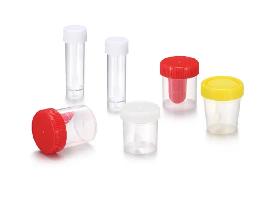
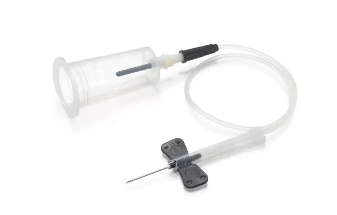
1. Sample Collection
Biological samples are taken with caution to prevent contamination.
2. Fixation and Preservation
To preserve structure, chemical agents are used to preserve specimens.
3. Slide Mounting Procedures
The samples are put on slides through standardized procedures to view them clearly.
4. Observation Staining Techniques
Contrast and cellular structures are enhanced by staining dyes.
5. Labelling and Cover Slipping
Last-minute preparation is to make sure that it is prepared safely and correctly identified.
Laboratory observation procedures in a scientific environment are based on these procedures.
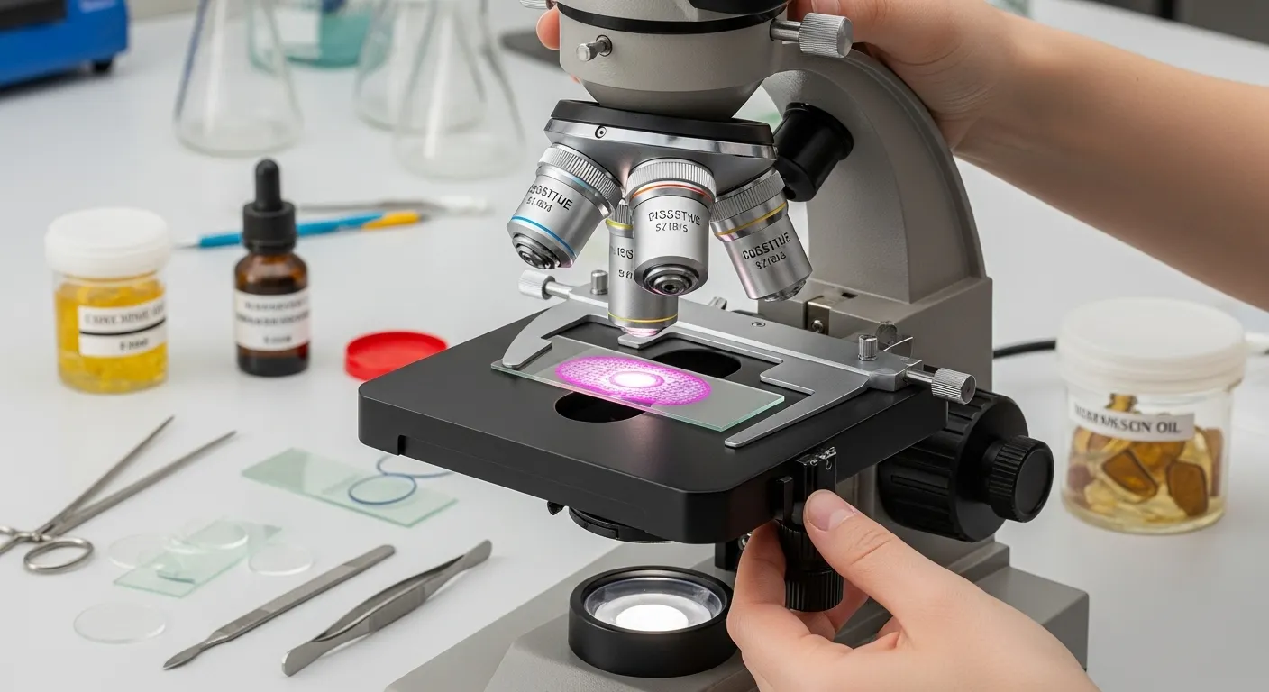
In the Laboratories, Microscope Tools and Equipment
The current microscope facilities in the lab are very crucial for proper analysis. Depending on the degree of detail, laboratories employ a mix of instruments.
Common Tools Include:
• Optical microscopes
• Electron microscopes
• Trinocular microscopes
• Digital imaging systems
• Magnification-based analysis tools
One of the most suggested alternatives to the professionals is the trinocular microscope because it can be used to view and record images in real-time. These tools allow the microscopic analysis of samples, the magnification of samples, and laboratory work with accuracy.
Microscope Slide Observation Methods and Techniques
Microscope slide observation techniques and training are important in the success of the methods. To make it clear and achieve the right outcomes, it is relevant to deal with it.
Effective Techniques Include:
• Adjusting magnification gradually
• Appropriate lighting settings.
• Use of immersion oil with high-resolution lenses.
• Calibration and use standards of microscopes.
• Following sample imaging workflow protocols
Advanced labs also use cytological examination techniques to examine individual cells and detect abnormalities. These methods enhance the proper microscopic examination and professional specimen viewing processes in studies.
Workflow Laboratory Microscopic Specimen Analysis
The efficiency of the results is identified by the workflow and laboratory specimen analysis tools.
An Average Workflow Comprises:
- Collection and labelling of samples.
- Preparation and staining
- Slide observation under the microscope
- Recording and documentation of images.
- Data interpretation
This is a typical clinical laboratory microscopy and biomedical specimen examination workflow.
The existing systems are a combination of digital imaging and biological specimen handling to improve accuracy and speed.
Scientific Investigations: Advanced Microscopy
Microscopy techniques are critical in scientific research studies that give profound information on cellular structures.
Common Advanced Techniques:
- Tissue analysis through histological analysis of the tissue sample.
- Cellular morphology: Study of cell structure.
- Microstructure examination to examine the biological aspects in detail.
- Disease research by biomedical imaging analysis.
- Scientific discovery, which is based on specimen research.
These procedures aid in the in-depth cellular study and enhance accurate diagnosis in laboratories.
Uses of Microscopic Specimen Observation
Microscopy is applied in various industries and research.
Major Applications Include:
- Healthcare diagnosis and illnesses.
- Microbiology research
- Pharmaceutical development
- Forensic investigations
- Environmental sample analysis
- Biomedical research studies
Applications of diagnostic microscopy in healthcare aid in the detection of infections, tissue abnormalities, and cellular changes in the early stages.
Typical Problems in the Microscopic Analysis
Despite the sophisticated equipment, labs have difficulties with the microscopic analysis of samples.
Common Issues Include:
• Poor slide preparation
• Incorrect staining techniques
• Low-quality microscope calibration
• Biological sample pollution.
• Improper magnification settings
In response to them, the laboratories follow strict laboratory observation rules and invest in the newest diagnostic microscopy devices.
The Reason to Trust Lab as Microscopy Lab Equipment
TrustLab China offers quality laboratory solutions, which are oriented towards accuracy and reliability. Their tools aid in fundamental and sophisticated microscopic examination of specimens in the laboratory.
Their Equipment Supports:
• High-precision biological imaging
• Improved visualization techniques of the specimen.
• Research-grade microscope analysis
• State-of-the-art microscopy systems within the lab.
Trustlab helps the global labs to deliver consistent and accurate outcomes via innovation and quality engineering.
Conclusion:
Microscopic Specimen Observation is more than just a laboratory process. It is an opening to the discovery of life in its simplest form. It helps to develop science in any direction, starting with diagnosing diseases and continuing with research.
The right tools, the right techniques, and the right laboratory systems allow scientists to get unparalleled clarity and accuracy in the analysis of biology.
Trustlab China can offer you a better understanding, more insights, and microscopy solutions at a professional level. Watch our new high-tech trinocular microscope systems at work and bring your lab work to a new level.