Updated 1 week ago
Histological Specimen Observation Guide | Techniques & Microscopy
sidrahafeez754
Histological Specimen Observation
Advanced Methods of Histological Specimen Observation in Medical Laboratories
One of the most significant bases of modern medical diagnostics and biological studies is the Histological Specimen Observation. It can be defined as the close examination of tissues using a microscope to learn about the structure of cells, disease patterns, and biological behavior on a microscopic scale. In simple terms, it is the science of observing things that are not visible to the naked eye.
In hospitals, research centers, and diagnostic laboratories, histology of specimens is very critical in the diagnosis of diseases like cancer, infection, and degenerative diseases. Scientists and pathologists analyze the morphology of tissues, cellular architecture, and microscopic tissue analysis using histology specimen analysis to draw the right conclusions.
The Knowledge of the Observation of Histological Specimens
The systematic microscopic examination of biological specimens is called Histological Specimen Observation. These samples can be biopsies, surgical, or laboratory experiments.
The Process Involves:
• Magnified structure examination of cells.
• Compound microscopes- histological analysis of slides.
• Interpretation of tissue specimen.
• Review of pathological specimens to detect disease.
The aim is to learn the behavior of tissues under normal and pathological conditions. The analysis of microscopic pathology is able to detect the smallest changes in cell structure.
This also involves histological pattern recognition and helps in the differentiation of normal and diseased tissues.
Processes and Workflow of the Histopathology Lab
To monitor the histological specimen, a good workflow of histopathology laboratory activities is required. The integrity of the tissue sample is guaranteed by each step.
Key Steps Include:
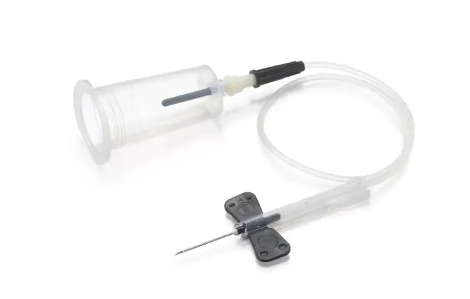
• Collection and Fixation of Tissues
Sample fixation and processing help to preserve the tissue structure with the help of such chemical solutions as formalin.
• Sectioning and Preparation of Tissues
Microtome section analysis gives thin tissue sections that are to be viewed under a microscope.
• Paraffin Embedding Process
Tissues are stabilized by parafilm or wax embedding to be cut.
• Slide Preparation Techniques
The steps involved in preparing microscope slides make sure that the tissue samples are easily mounted.
• Staining Evaluation
Histological staining methods accentuate cellular details.
All these measures assist in laboratory specimen handling processes, and there should be no distortion in the process.
Role in Viewing Histological Specimens
Tissue embedding is one of the most important steps in the observation of histological specimens. Unless correctly embedded, precise microscopic tissue analysis is challenging.
Embedding helps in maintaining the tissue structure and allows thin sectioning to study the histological slides. The laboratory embedding cassette is one of the most reliable products that are involved in the same.
The Embedding Cassette's Role is in:
- Contain tissues during processing.
- Simple identification and labelling of specimens.
- Smooth paraffin-embedded specimen analysis.
- Better structural conservation of microscopic tissue analysis.
These cassettes make sure that the morphology analysis of tissues is not lost during the histopathology laboratory process.
Better Observation with the Assistance of the Histological Staining Techniques
Staining is the observation of a histological specimen. No cellular details can be observed under the light microscopy without staining.
The Most Common Staining Techniques are:
- Hematoxylin and Eosin (H&E) Staining
The most popular technique of clinical histology evaluation.
- Special Stains of Tissues
Applied in the detection of bacteria, proteins, and cellular components.
- Immunohistochemical Staining
Assists in the diagnosis of disease tissue through the detection of particular antigens.
These methods enhance the interpretation of microscopic images and aid in the analysis of biomedical specimens.
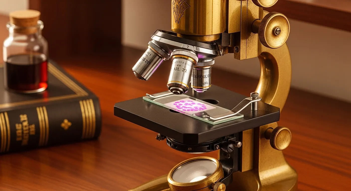
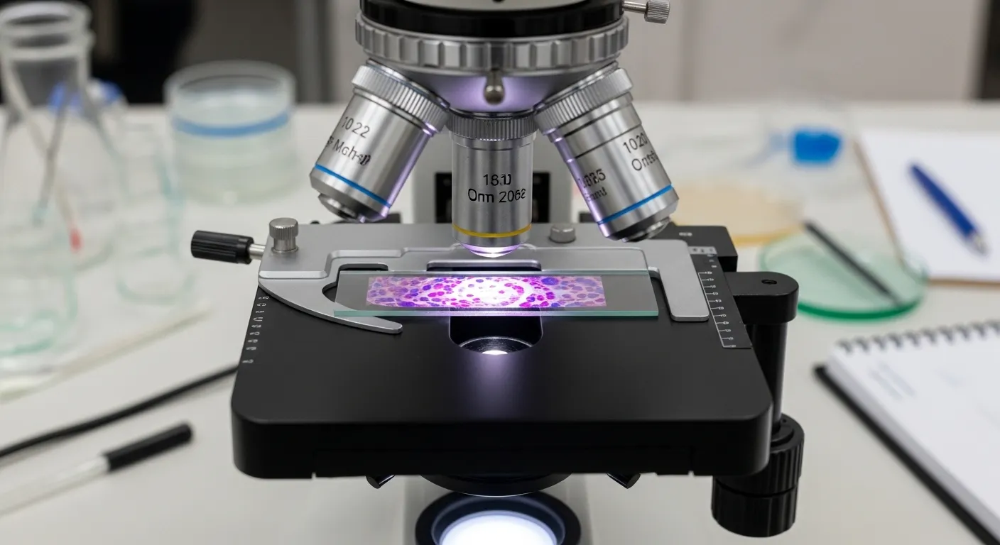
Microscopy Methods in the Examination of Histological Samples
The primary tool of histological observation in the microscopy procedure is the microscope. Different levels of detail are provided by different microscopy techniques.
The Important Microscopy Techniques are:
- General study with a compound microscope.
- A high magnification tissue study to study the structure of the cells in detail.
- Slide-based observation methods of regular diagnostics.
- State-of-the-art digital histology images.
- Tissue analysis electron microscopy: ultra-structural studies.
These methods allow scientists to perform microscopic examination of tissues in an extremely precise and clear way.
Clinical Significance of Histological Examination of Specimen
The histological specimens are directly observed in the clinical setup, which is directly linked to the diagnosis and treatment planning of the disease.
It Supports:
- Clinical histology diagnosis of a patient.
- Diagnosis of cancer with biopsy samples.
- Diagnostic tissue tests of diseases and infections.
- Histopathological interpretation of medical reports.
Doctors can detect tissue structure and functional abnormalities through cellular pathology evaluation. This renders the examination of histology specimens as one of the most dependable diagnostic tools in contemporary medicine.
Analysis of Histological Specimens in a Step-By-Step Process
The accuracy of the observation of the histological specimen is achieved in a systematic fashion.
Step 1: Tissue Collection
When collecting biopsy or surgical samples, care is exercised.
Step 2: Fixation
Tissues are preserved using chemical fixatives.
Step 3: Embedding
Embedding of tissues in paraffin is through the embedding of cassettes.
Step 4: Sectioning
Microtomes are used to cut thin slices of tissue.
Step 5: Staining
To contrast the slides, they are stained.
Step 6: Microscopic Observation
Microscopes are used to analyze histological slides.
Step 7: Interpretation
Experts diagnose it using methods of interpretation of histological data.
Best Practices in the Observation of Histological Specimens
Laboratories are informed by stringent regulations to achieve the right results.
Best Practices Include:
• Correct fixation and processing of samples.
• Making good microscope slides.
• Seeing clean laboratory handling practices.
• Correct sectioning and preparation of tissues.
• Following standardized histopathology laboratory procedures
The practices also enhance consistency in the study of microscopic pathology and minimize errors in diagnosis.
Advanced Applications in the Study of Histological Specimens
The extended uses in modern science have been in the advanced fields of histology.
These Include:
- Analysis of morphological variation of studies.
- Genetic research of structural tissue organization.
- Disease progression monitoring
- Drug development: The development of drugs by the analysis of biomedical specimens.
- Histological pattern recognition in AI-aided diagnostics.
These applications provide value to microscopic tissue analyses in research and healthcare.
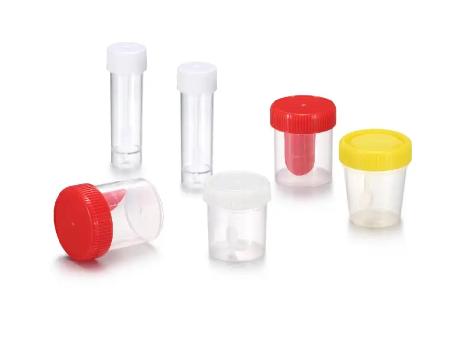
Importance of Laboratory Aids in Histology
Laboratory equipment is necessary to make the appropriate observation of histological specimens.
Key Tools Include:
- Microtomes for sectioning
- Preparation of staining jars to prepare slides.
- Introduction of cassettes to treat tissues.
- Observation compound microscopes.
These devices guarantee the successful systematization of the work of histopathology laboratories and enhance the accuracy of diagnoses.
Conclusion:
Histological Specimen is not a simple laboratory procedure to observe; it is the basis of the modern diagnosis of diseases and biological knowledge. Each of the steps taken since the fixation of tissues to the microscopic study of tissues is a step towards a deeper understanding of cellular life. Laboratories can be more accurate and clear than ever before with the new tools, such as embedding cassettes and better methods of histological staining.
Should you require the answer to the dilemma of credible lab solutions that will help to examine the histology specimen correctly, Trustlab China can provide you with high-quality tools that are professionally used and may be considered scientifically high-quality.