Updated 2 months ago
Specimen Imaging Techniques in Modern Laboratories
sidrahafeez754

Specimen Imaging Techniques
Cutting-Edge Specimen Imaging Techniques for Healthcare Professionals
The current science and healthcare known as specimen imaging techniques is a power that can enable us to learn what the human eye cannot see. These techniques help scientists, pathologists, and researchers to see biological systems clearly and accurately, whether it is small cells or a complicated tissue structure.
Imaging of biological specimens is no longer just an act of observing today. It has evolved into an extremely advanced field incorporating optics, digital systems, and computing equipment. The new ways through which a laboratory diagnoses a disease, investigates an infection, and analyzes a cell structure are through clinical specimen imaging and lab specimen imaging.
What are Specimen Imaging Techniques?
Specimen imaging techniques are a collection of scientific processes that are used to examine biological specimens such as tissues, blood, bacteria, and cells. These methods include:
- Imaging techniques of microscopic specimens.
- Electronic imaging of specimens.
- Imaging of histological samples.
- Intermediated imaging of samples.
- Specimen-based imaging analysis
Basically, these techniques enable specialists to perform research and diagnosis of biological samples and tissue visualization procedures.
They find wide use in:
• Hospitals
• Pathology laboratories
• Biomedical research centers
• Clinical diagnostic labs
The Importance of Specimen Imaging in Medical Diagnostics
The imaging of clinical specimens is of utmost importance. It helps directly in detecting and planning the treatment of disease and research development.
Key benefits include:
- This is because it early detects ailments like cancer.
- Normal morphology of the cells.
- The improved medical lab diagnostics.
- Very high accuracy of imaging pathological samples.
- Better understanding of tissue changes.
In the present-day healthcare, observation is not imaging; it is imaging-based disease detection that allows the doctor to make life-saving decisions in a shorter period.
Lab Workflow and Imaging Procedures
Imaging processes in the laboratory will also help in understanding how the samples can be taken through the collection process to analysis.
A typical working process entails:
- Biological sample sources.
- Slide preparation with staining.
- Slide-based imaging procedures.
- Microscopy or imaging.
- Digitalization of high-resolution devices.
- Examination of specimen pictures using software.
- Storage of images that can be used in the future for diagnostic purposes.
Such a systematic nature ensures the accuracy and reliability of every process of the analysis.
Significant Specimen Imaging Technologies
In modern laboratories, the imaging techniques for specimens are based on different technologies and are implemented to fulfill a specific diagnostic task.
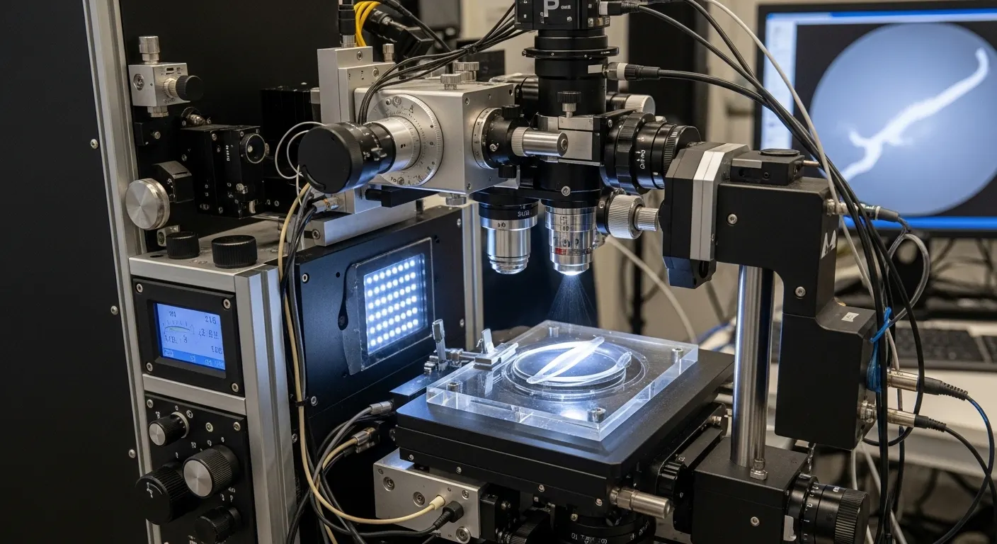
Microscopic Imaging Techniques
Microscopy continues to underlie biological research. Researchers are able to observe the cells and tissues in detail with good optical systems. The systems are used to support the process of microscopic imaging and high-resolution imaging of specimens to achieve precise results.
Fluorescence Microscopy Imaging
This method makes use of fluorescent dyes to demonstrate structures in specimens. It finds wide use in:
- Cancer research
- Genetic studies
- Protein localization
- Confocal Biological Imaging.
Confocal microscopy allows the images to be 3D-like as the out-of-focus light is eliminated. It is needed in cellular imaging methodologies and deep tissue studies.
Electron Microscopy Visualization
The electron microscopes are very high in magnification and therefore enable the scientists to observe the sub-cellular structures.
This supports:
• Virus imaging
• Molecular studies
• Structural biology visualization
DAVP Digital Imaging Systems
Present-day laboratories apply AI-based applications that are used to visualize and analyze digital specimens.
Specimen Imaging CT, MRI
They are sophisticated diagnostic imaging processes used on internal biological structures and bigger specimen observations.
Biological Specimen Imaging in Health Care
Biological specimen imaging can be used in numerous sciences.
Pathology
• Histopathological imaging techniques
• Disease classification
• Tumor analysis
Microbiology
• Bacterial imaging
• Virus structure observation
• Infection diagnosis
Cancer Research
• Cancer specimen imaging.
• Tumor growth tracking
• Cellular mutation detection
Biomedical Research
• Drug testing studies
• Genetic research
• Molecular behavior analysis
Biomedical imaging and specimen examination procedures are greatly relied upon in the applications.
AI in Specimen Imaging and Digital Pathology
The digital revolution has changed the way laboratories are operated.
Modern systems have turned into:
• AI-based image recognition
• Automated tissue scanning
• Cloud-based storage systems
• Real-time collaboration tools
This facilitates the digital pathology imaging systems and improves accuracy in laboratory diagnostic imaging. AI also enhances the analysis of the imaging of the specimen and is faster and more dependable in diagnosing.
Analysis and Interpretation of Specimen Imaging
This includes:
- Specimen analysis techniques
- Tissue structure pattern recognition.
- Cell morphology analysis
- Comparative disease studies
- Quantitative image measurement
Raw images are converted into valuable clinical information using state-of-the-art software. This is required to diagnose imaging of specimens and research validation.
Advantages of Modern Specimen Imaging Techniques
The new imaging systems possess several advantages:
• Good diagnostic accuracy.
• Fast data processing
• Better disease visualization
• Better visualization of the tissue structure.
• Reliable research outcomes
• Improved laboratory efficiency
The benefits have seen laboratory imaging processes to be superior and more reliable than ever.
Trustlab-China Solutions of Specimen Imaging
Trustlab is a state-of-the-art laboratory solution that will be used to enable new optical microscope specimen imaging methodologies with accuracy and reliability.
Our microscope systems are optical and provide:
• High clarity imaging
• Stable magnification control
• Advanced lens systems
• The best use of microscopic imaging of specimens.
Most hospitals, universities, and diagnostic laboratories have the tools to analyze biological samples and carry out research based on imaging.
Conclusion
The world of specimen imaging techniques is transforming at a faster rate than ever. Every new step in the history of microscopy, up to the modern AI-powered digital technology, has led science to the expected level of perfect accuracy.
The contemporary laboratories are founded on the most recent medical imaging of specimens, histological imaging analysis, and biomedical specimen imaging applications to discover about life at the tiniest level.
Innovation is paired with precision at Trustlab-China, which offers the tools that will change the perspective on biology. Once you are ready to improve your experience in the laboratory, think about the newest optical solutions and leap into the future of imaging without any problems.
Visit Trustlab-China and see how modern specimen imaging can transform your diagnostic and research.
Related Articles

Does a Gynecological Exam Hurt? The Design Science Behind Patient-Friendly "Swabs for Women"
From Sample Preparation to Storage: How Screw Cap Centrifuge Tubes Optimize Daily Lab Workflows