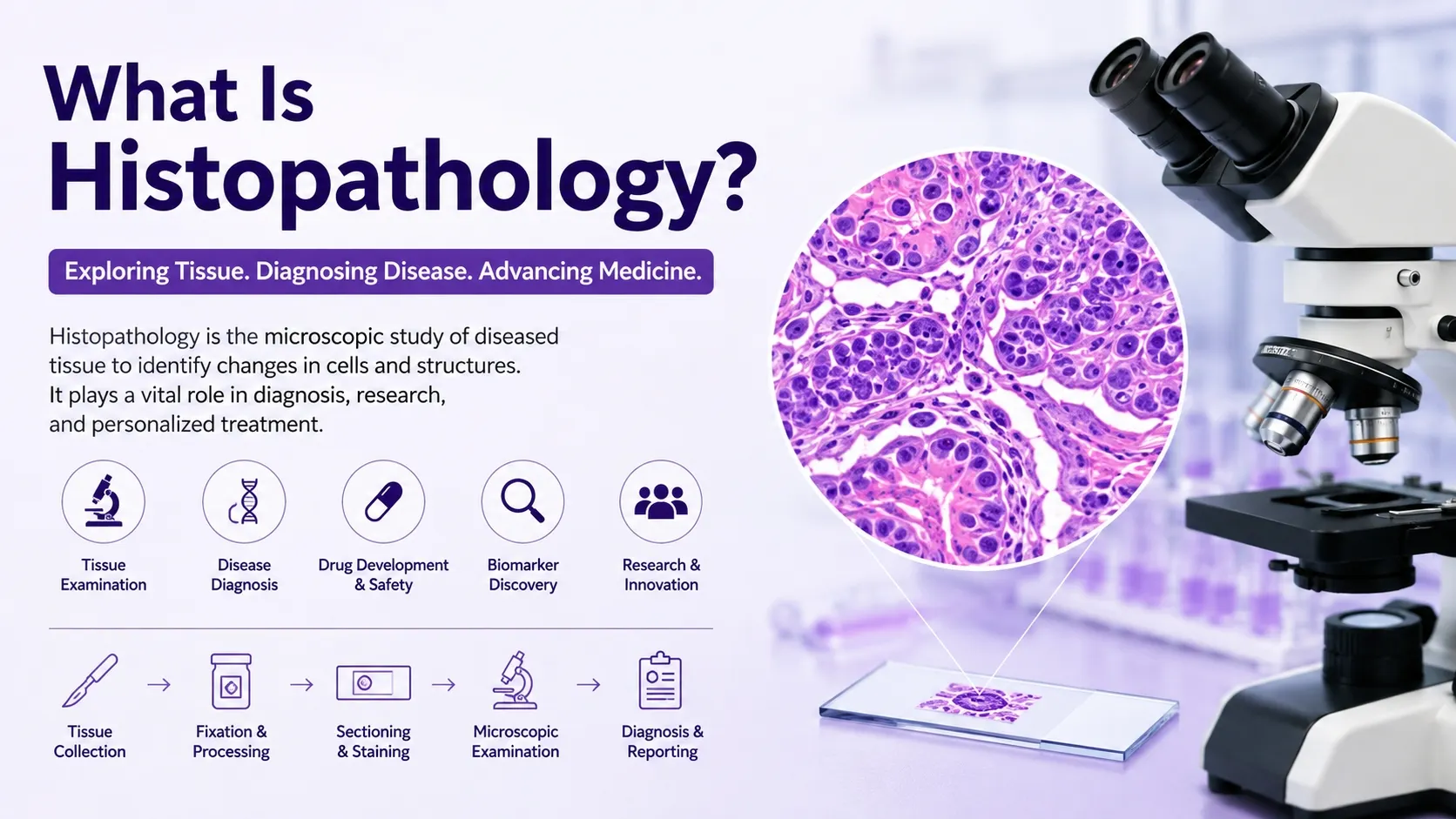
what is histopathological
Histopathology is among the most crucial tools in contemporary medicine and research in biomedical sciences. A pathologist and researcher can use a microscope to look at the tissue and determine what changes have occurred at the level of the cell and structure that may relate to the disease. Whether it is determining the nature of the disease in cancer diagnosis or aiding in the development of drugs, histopathology plays a crucial role in understanding the impact of a disease on organs and tissues.
The field of histopathology is increasingly vital in the era of digital pathology, artificial intelligence and spatial biology, and is playing an increasingly critical role in the global clinical, biotech and academic community.
What Is Histopathology?
Histopathology: The science of examining tissues under the microscope. It includes the interpretation of stained tissue sections to look for abnormalities in cells, architecture of the tissue, and structure of the organs. Histopathology is valuable for pathologists to find out if the tissue is normal or if it is damaged, due to diseases such as inflammation, infection, fibrosis, and tumors.
Histopathology retains the organization of tissues as opposed to cytology which considers isolated cells. This enables scientists to study the interactions between cells in their native habitat, particularly in the diagnosis of cancer and in the understanding of the progression of disease.
Histopathology is commonly used in:
- Cancer diagnosis and tumor grading
- Toxicology and drug safety studies
- Biomarker discovery
- Translational medicine
- Infectious disease research
- Preclinical pharmaceutical studies
- Academic and clinical pathology research
Digital pathology systems are now integrated with traditional microscopes in modern labs to facilitate collaboration, remote review, and image analysis processes.
Histology vs. Histopathology
Although the terms are often confused, histology and histopathology are different disciplines.
Histology is a study of normal tissue structure and anatomy. It sets a standard for the healthy appearance of the organs and cells.
Histopathology involves the study of abnormal tissue and is the study of the structural or cellular abnormalities that are the result of disease.
| Feature | Histology | Histopathology |
|---|---|---|
| Main Focus | Healthy tissue structure | Diseased tissue analysis |
| Purpose | Study normal anatomy | Detect pathological changes |
| Applications | Education and baseline research | Disease diagnosis and research |
| Typical Findings | Normal cell arrangement | Tumors, inflammation, necrosis |
Understanding this distinction is important for researchers selecting appropriate tissue analysis methods for their studies.
The Histopathology Workflow
The following steps are some of the basic steps in a histopathology workflow that are carefully controlled to preserve the integrity of the tissue and to yield accurate microscopic results.
1. Tissue Collection
The initial step is the collection of tissue by biopsy, surgery, and/or experimental sampling. Handling at this stage is important as the tissue may start to degrade and cause a problem in the diagnosis.
2. Tissue Fixation
Chemical fixation, like formalin, is used to collect the tissue. Fixation stops tissue breakdown and maintains cell shape. In a pathology lab, formalin-fixed paraffin-embedded (FFPE) tissue is a routine technique for preparing samples.
3. Tissue Processing and Embedding
Tissue fixation involves removing water from tissue. The specimen is then fixed in paraffin wax so as to form a stable block from which sections can be cut.
4. Sectioning
The material is cut to a thickness of 4–5 micrometers with a microtome to produce thin tissue slices. These sections are placed on the microscope slides and stained and examined.
5. Tissue Staining
Staining provides contrast of the tissues and shows the cellular structure. The various staining techniques depend on the diagnostic or research goals.
6. Microscopic Evaluation
The stained slides are examined by pathologists or trained scientists under a microscope or digital pathology system, to look for abnormalities.
7. Interpretation and Reporting
This final step is to document observations, diagnoses and pathological findings in a pathology report.
Common Histopathology Staining Techniques
Hematoxylin and Eosin (H&E) Staining
The "gold standard" in histopathology is H&E staining. Hematoxylin will stain nucleus blue/purple and eosin will stain the cytoplasm and extracellular structures pink. This stain can be used to give an overview of tissue architecture and cellular morphology.
Immunohistochemistry (IHC)
IHC is a technique that detects particular proteins in tissue sections using antibodies. It is a commonly used in cancer research, biomarker analysis and precision medicine applications.
Special Stains
Special stains are used to distinguish special tissue components or microorganisms. Common examples include:
- PAS stain for carbohydrates and mucins
- Masson’s Trichrome for collagen and fibrosis
- Gram stain for bacteria
- Prussian Blue for iron deposits
Immunofluorescence (IF)
Immunofluorescence is a technique that takes advantage of fluorescent dyes to make proteins or molecular targets visible in tissues. Multiplex IF enables the analysis of several biomarkers at the same time.
Why Histopathology Is Important
Histopathology is still important to the present day because it brings together molecular biology and the real tissue structure and the behavior of the disease.
Cancer Diagnosis
Histopathology has a key role in the detection, classification, staging and margin assessment of cancer. Tissue morphology is analysed to diagnose malignancy and to facilitate treatment planning.
Drug Development and Toxicology
Histopathology is used by pharmaceutical and biotech companies to assess organ damage, treatment effect and tissue toxicity in preclinical studies.
Biomarker Discovery
In oncology, immunology and translational medicine, advanced histopathology techniques assist in the identification and validation of biomarkers.
Personalized Medicine
Personalized treatment strategies are possible with the help of histopathology and molecular diagnostics using tissue-specific biomarkers and protein expression patterns.
Biomedical Research
Histopathology is used in research for study of disease mechanisms, tissue regeneration, inflammation, fibrosis and cellular interactions in experimental models.
Digital Pathology and AI in Histopathology
A revolution that has taken place in the field of pathology is the conversion of glass slides to high resolution digital images known as digital pathology. Whole slide imaging (WSI) systems enable remote collaboration, storage in the cloud, and computational analysis.
Histopathology workflows are now increasingly incorporating AI and machine learning. AI can help with:
- Cell counting
- Tumor segmentation
- Biomarker quantification
- Pattern recognition
- Predictive pathology analytics
The field of computational pathology is growing exponentially in most areas of study, particularly in graph-based tissue analysis and automated diagnosis and classification systems.
Challenges in Histopathology
Although significant progress has been made, there are still some limitations on the use of histopathology.
Sample Quality Variability
Slides may be of poor quality and may not be diagnostically accurate due to poor fixation, damage to tissues, or inconsistent staining.
Interpretation Complexity
Highly trained specialists are needed to interpret the histopathological findings. Several diseases exhibit similar morphology, and are difficult to diagnose.
Data Management
Digital pathology generates very large image files which necessitate advanced storage and computational infrastructure.
Standardization
However, it is still a challenge to maintain reproducible staining and imaging protocols between different laboratories in various countries around the world for reproductive research studies.
The Future of Histopathology
Digital transformation and precision medicine are intrinsically linked with the future of histopathology.
Emerging technologies include:
- Spatial transcriptomics
- Multiplex imaging
- AI-assisted diagnostics
- Cloud-based pathology platforms
- Automated image analysis
- High-throughput tissue phenotyping
The innovations will enhance the speed of diagnosis, the reproducibility of diagnostics, and the scalability of diagnostics in clinical and research settings.
With the increasing complexity of computational pathology, histopathology will further transition from being a mainly visual to being a data-driven scientific platform.
Conclusion
Histopathology is still an integral part of the modern medical, pathological and biomedical research. It shows minute changes in the tissues that can be related to disease, aids drug development and scientific discovery.
Histopathology has seen the introduction of various new technologies that enhance its capabilities, precision, and effectiveness, ranging from classic H&E staining to AI-driven digital pathology. Histopathology is invaluable in the understanding of disease processes and tissue biology, for diagnosis of cancer, toxicology and in the context of translational medicine.