


Updated 2 months ago
NA 1.25 Condenser in Laboratory Microscopes for Light Control

Admin

Laboratory Microscopes
Clear microscope images depend on more than objective magnification. In routine biological observation, illumination angle, condenser alignment, and aperture adjustment all influence image contrast, resolution, and depth of field.

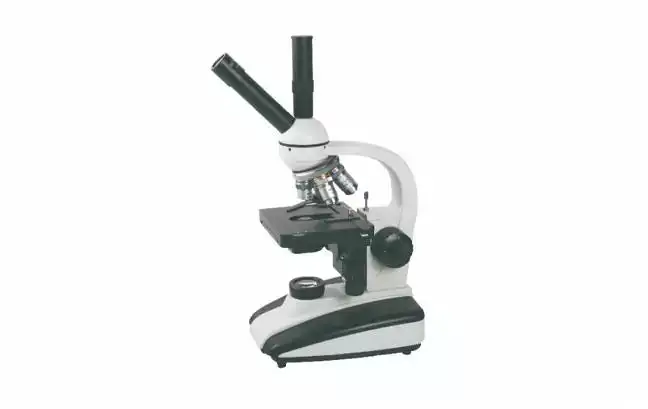
In many Laboratory Microscopes, these functions are managed through an NA 1.25 Abbe condenser with an integrated iris diaphragm. This optical system, when properly adjusted, demonstrates a controlled cone of light delivery system that helps users illuminate fine structures in the specimen without imprecise glare and contrast.
This article explores the collaboration of the condenser and iris diaphragm and describes how Trustlab integrates customisable illumination in its Laboratory Microscopes.
What Is an NA 1.25 Abbe Condenser?
This type of condenser is found under the stage of a microscope. It gathers light emitted from the illuminator and focuses it on the plane of the specimen.
Numerical aperture, or NA, is a measurement of the condenser's ability to propagate light at an angle. An NA 1.25 Abbe condenser is adequate for standard low-power observation and high-resolution work with oil-immersion objectives.
This type of condenser performs multiple functions:
1. Light Concentration: Specially designed to evenly concentrate light.
2. Resolution Assistance: Produces an even larger cone of light which helps resolve very fine details.
3. Objective Compatibility: Modifies the illumination aperture for objectives with a wide range of numerical apertures.
4. Oil-Immersion Observation: Provides very high-NA observation with the use of immersion oil between the condenser lens and the microscope slide.
5. Field Uniformity: Reduces uneven brightness in the viewing field.
When used dry with 10X or 40X objectives, the condenser operates at a lower effective NA. For 100X oil-immersion observation, oil coupling can reduce light refraction and allow the system to use more of the condenser's available aperture.
How the Iris Diaphragm Controls Illumination
The iris diaphragm is an adjustable aperture built into the condenser. It changes the diameter and angular width of the light cone entering the objective.
The iris diaphragm should not be treated simply as a brightness control. It primarily serves to control the aperture of illumination.
Changing it influences three key attributes of the image in laboratory microscopes:
Contrast
Reducing the iris diaphragm helps control stray and oblique illumination. This, in turn, helps in viewing somewhat transparent and lowly contrasted specimen features.
Resolution
Increasing the diaphragm allows for the use of a condenser of a larger numerical aperture (NA) and allows the objective to work with the illumination of wide angles. This supports higher resolution, although excessive opening may produce a bright image with weak contrast.
Depth of Field
Closing the diaphragm increases the apparent depth of field. More of a thick specimen may appear focused at the same time, but fine-detail resolution will be reduced.
Comparing Iris Diaphragm Settings
The most useful setting is usually between fully open and heavily closed. A practical starting point is to adjust the condenser aperture to approximately 70–80% of the objective's numerical aperture.
| Iris setting | Image characteristics | Main limitation | Typical use |
| Fully open | Maximum illumination aperture and potential resolution | Lower contrast and brighter background | Dense stains or high-resolution observation |
| Moderately open | Balanced contrast, brightness, and detail | Requires adjustment when changing objectives | Routine stained slides and biological smears |
| Partially closed | Increased contrast and depth of field | Reduced fine-detail resolution | Transparent or lightly stained specimens |
| Heavily closed | Strong edge contrast and darker background | Diffraction artifacts and loss of resolution | Initial focusing or coarse inspection |
The diaphragm setting should be reviewed whenever the objective is changed. A setting suitable for a 10X objective will generally not provide the best results with a 40X or 100X objective.
Adjustment Procedure Based on Application
The overwhelming majority of general designs of models of microscopes have similar features, yet some specific designs are necessary for the different applications. Furthermore, some small adjustments may be necessary for optimisation and for achieving repeatable results.
1. Choosing Microscope Objective for First Focus
Place the specimen on the stage. Choose a suitable objective for a first, rough focus, turn on the microscope and adjust the light intensity as per your requirement.
2. Aligning Optical Axis and Cone of Light
The condenser is kept below the slide on the stage. The cone of light should be adjusted to the optical axis by centering screws.
3. Start With the Iris Open
Keep the iris diaphragm of the condenser in the fully opened position. Then, slow closing of the diaphragm should be done in steps while viewing the specimen.
4. Contrast and Resolution
The resolution of the specimen should be adequate in order to delineate the boundaries of the specimen and to be able to distinguish the finer details of the specimen. if the iris diaphragm is closed too much, the image will be dark and of poor resolution.
5. Adjust Brightness Independently
Use the lamp-intensity control or neutral density filters to adjust brightness. Do not reduce illumination by closing the iris too far, as it will diminish resolution.
6. Recheck after changing magnification
Each objective has a different numerical aperture. Condenser height, iris opening, focus, and lamp intensity may all require minor readjustment.
Typical Illumination Issues
Problems with illumination are often the result of improper setting of the condenser rather than contamination of optics or improper specimen preparation.
•Bright image with poor detail: The iris diaphragm may be too open.
•Dark or grainy image: The diaphragm may be too closed.
•Uneven field illumination: The condenser could be off-centre or in the wrong position.
•Reduced oil-immersion resolution: The oil may be contaminated, absent, or poorly distributed.
•Halos around structures: The aperture of the condenser may be too closed.
•Inconsistent contrast: The diaphragm and/or the condenser may be defective.
Systematic adjustment is generally more effective than increasing lamp brightness alone.
Trustlab's Approach to Laboratory Microscope Illumination
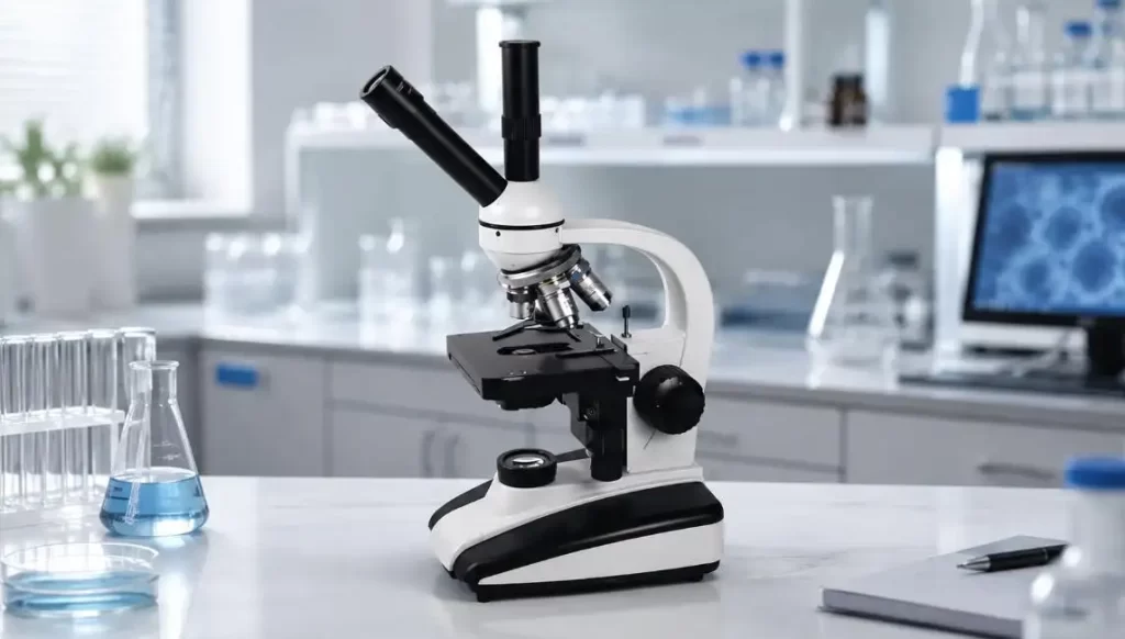
Trustlab constructs Laboratory Microscopes emphasizing optical alignment and ease of use for daily operations. The illumination system for the microscopes in use for biological observation is designed using a halogen light coupled with an NA 1.25 Abbe condenser and an adjustable iris diaphragm.
The principal design components comprise the following:
•Halogen light (6V/20W): Provides an adjustable light intensity.
•Smooth diaphragm: Allows for continuous variations of the diaphragm opening.
•Double-layer mechanical stage: Facilitates the best positioning of the specimen for observation.
•45° inclined and 360° binocular viewing head: Made for use by multiple individuals.
•Acromatic objectives: Allow the observation of 40X to 1000X of total magnification.
•WF10X eyepieces: Allow a viewing field adequate for instruction and for routine laboratory use.
The combination of these features allows the user to gain optimal observation for all of their needs.
The Importance of Accurate Control of Illuminating Intensity
Accurate control of illuminating intensity assists in the observation of the outlines of cells, of nuclei and cytoplasm, of microorganisms, of microorganisms in the stained tissue, and of other tissues. It will enable the user to obtain the best possible image of the specimen without disturbing the specimen for additional staining.
Well-adjusted Laboratory Microscopes are ideal for educational laboratories, clinical training, and routine biological analysis, as they provide:
•More consistent image quality
•Faster focusing
•Enhanced visibility of low-contrast structures
•Reduced eye fatigue
•Increased repeatability of observations
To Conclude
The NA 1.25 Abbe condenser with iris diaphragm incorporated in Laboratory Microscopes offer excellent control in the illumination system. The condenser focuses specimen illumination, and the diaphragm controls the angle of illumination that pertains to the objective.
Condenser adjustments and lamp intensity, alignment and aperture adjustments can maximise contrast and minimise resolution reduction. Trustlab has incorporated all of these to aid the user in obtaining clear and reproducible observations in standard lab conditions. Stable halogen lighting, adjustable condensers, stable micrometric stages, and effective viewing mechanisms are incorporated in its Laboratory Microscopes.
FAQs
Q1. What is the function of an NA 1.25 condenser?
It helps in the projection of a large cone of light onto the specimen, thereby aiding in observation of the specimen in great detail.
Q2. What does the iris diaphragm affect?
It controls light which affects the resolution and the depth of field.
Q3. Is the diaphragm best when fully open?
Not necessarily. A better compromise of contrast and resolution is usually found with a diaphragm that is not fully open.
Q4. How best can the condenser be set for a 100X objective?
The condenser should be raised close to the slide, and immersion oil should be used.
Q5. Why is the image bright but has low contrast?
This is usually due to a diaphragm that has been set to a too much open position, which allows too much light to the objective.