Updated 2 months ago
Specimen Slide Microscopy Guide: Preparation, and Lab Analysis
sidrahafeez754

Specimen Slide Microscopy
Enhancing Tissue Resolution Through Optimized Specimen Slide Microscopy Staining Protocols
The modern biological research and clinical diagnostics center on Specimen Slide Microscopy. It renders the unseen seen. This technique allows scientists to examine life at a cellular level with accuracy and clarity, from the examination of microscopic slides to the advanced examination of specimen slides.
As a student, researcher, or laboratory professional, knowing about sample slide microscopy is the gateway to dependable findings, improved diagnosis, and scientific breakthroughs.
What is Specimen Slid Microscopy?
The term Specimen Slide Microscopy is used to refer to the procedure of looking at biological samples placed on glass slides using a microscope. It entails the microscopic examination of samples using slides, the evaluation of microscopic samples, and the examination of biological slides under controlled conditions in the laboratory.
This method is widely used in:
• Medical diagnostics
• Biological research
• Labs of histology and cytology.
• Educational laboratories
It allows the visualization of laboratory specimens and the microscopic study of slides in detail.
Specimen Slide Microscopy is an Important Part of Labs
Accuracy is important in any lab. The tiniest error in the procedure of the specimen slide can affect the outcomes. This is the reason why microscopy of laboratory slides is significant in giving accuracy.
This is because it is needed:
• Helps in the observation of cell structure.
• Helps in clinical slide analysis.
• Early diagnosis of diseases.
• Improves biological sample visualization
• Guarantees good slide-based biological analysis.
When properly performed, it improves the quality of observation of the microscopic specimen and research results.
Types of Microscope Specimen Slides
Knowing the various microscope specimen slides assists you in selecting the appropriate method of analysis.
1. Prepared Microscope Slides
They are ready-to-use slides, which are typically used in education and research.
- Ideal for beginners
- Typically applied in teaching slide microscopy.
- Available in kits for students
2. Temporary Slides
- Prepared in a short time to be viewed.
- Used in quick viewing of microscope slides.
- Appropriate for live samples.
3. Permanent Slides
Produced by using sophisticated specimen preservation methods on slides.
- Long-lasting
- Important to research archives.
- Applications: Microscopy of histology slides.
Slides Preparation Step-by-Step Preparation of Slides
Good microscopy is founded on the making of a good slide. Follow these instructions for microscopy on a slide:
1. Sample Collection
Get a clean biological sample. This is the initial step in handling the specimen and preparing the slide.
2. Fixation
Preserve biological samples with chemicals. This maintains form and avoids deterioration.
3. Sectioning
Prepare the microtome sectioning procedure to get thin tissue sections to be utilized in the microscopy of tissue sections.
4. Mounting
Properly mount glass slides and apply proper coverslips.
5. Staining
Apply staining of microscopy slides to highlight structures. Stain the slide as per the standard procedures.
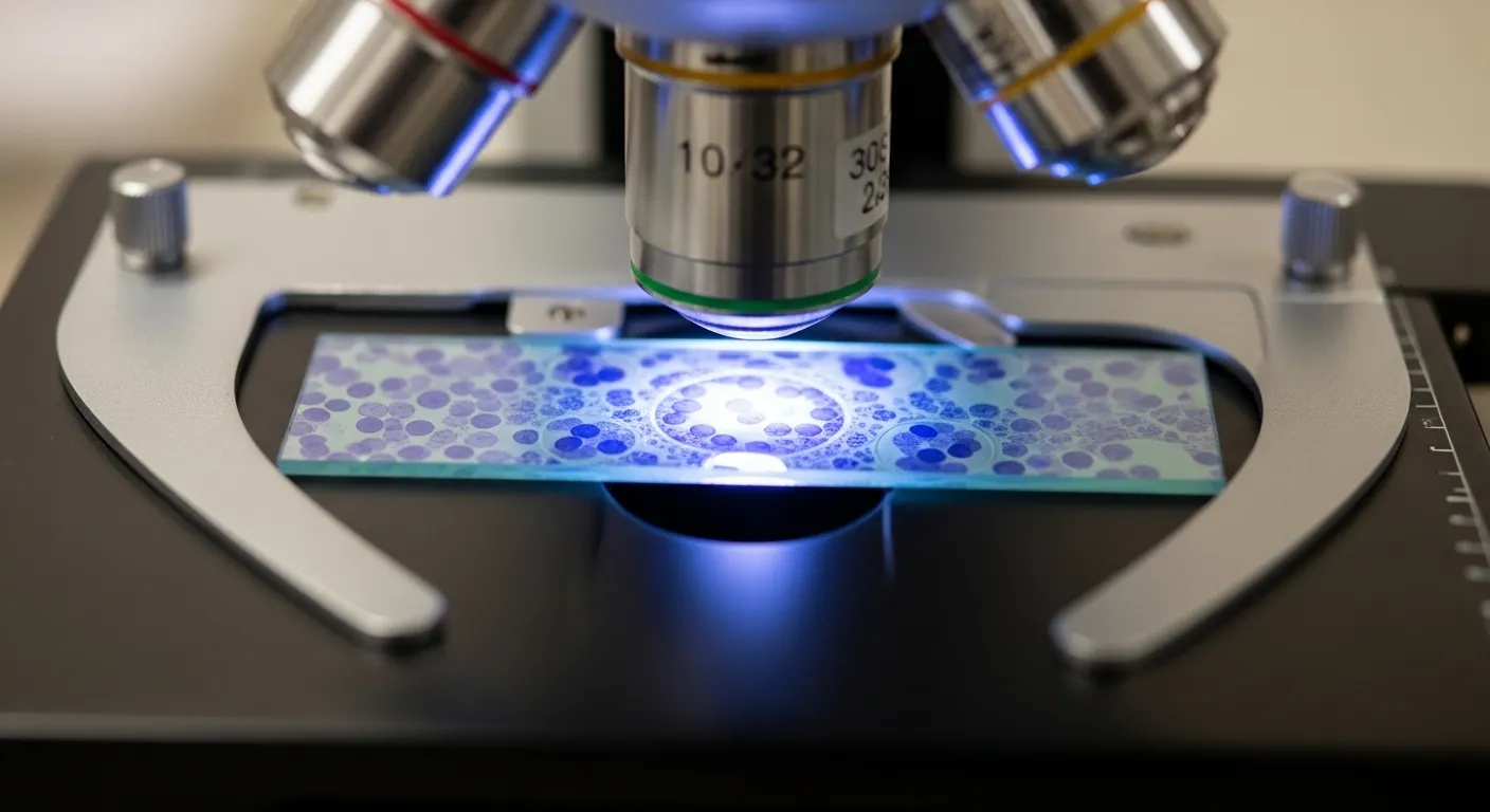
6. Observation
Biological slides are microscopically analyzed by placing the slide under a microscope.
Optimal Methods of Slide Microscopy of Specimens
Clarity and accuracy are improved by the choice of suitable slide microscopy techniques.
Light Microscope Slide Analysis
- Most common method
- Proper for standard optical microscopy slides.
- Applications: In schools and labs.
Compound Microscope Observation
- Offers higher magnification
- Excellent in microscope slide techniques.
Phase-Contrast Slide Microscopy
- Stainless contrast enhancement.
- Applicable to live cell observation.
Fluorescence Slide Imaging
- Uses fluorescent dyes
- Most appropriate for advanced sample imaging techniques.
Digital Slide Microscopy
- Allows viewing of specimens at high resolution.
- Remote analysis and storage.
Slide Microscopy Lab Methods
Laboratory microscopy is a methodology that is followed in contemporary laboratories in order to bring uniformity.
Key methods include:
• Managed workflow of specimen observation.
• Standardized slide handling in laboratories
• Good optical imaging of specimens.
• Microscope slide analysis in the laboratory.
These techniques enhance slide microscopy, which is based on research and provides reliable results.
Slide Microscopy of Histology and Cytology
Slide microscopy plays a crucial role in two significant scientific areas:
Histology Slide Analysis
Focuses on tissues. It involves:
• Histology slide examination
• Sectioning and staining of tissues.
• Laboratory analysis of biological slides in detail.
Cytology Specimen Analysis
Focuses on cells. It includes:
• Cytology specimen slides preparation
• Cellular abnormalities detection
• Clinical diagnostics
Both sciences are based on the accurate observation of the specimen on a slide.
Microscopic Observation of Slides
To amateurs, good observation practices are important.
Just do the following:
• Start with low magnification.
• Adjust focus slowly
• Use proper lighting
• Do not touch the surface of the slide.
• Maintain clean lenses
This enhances the observation techniques of microscope slides and guarantees clarity.
Using the Right Optical Microscope
A high-quality microscope is a large disparity in the microscopy of specimen slides.
In order to get professional outcomes, think more advanced solutions, like the ones provided at TrustLab.
These microscopes support:
• Precision light microscopy specimen slides examination.
• Improved use of compound microscope slides.
• Good biological slide microscopy.
• Appropriate microscopic analysis of the slides in the clinic.
The Microscope Slides are Used by Students and Professionals
The choice of the appropriate slides enhances learning and outcomes.
Look for:
• High-quality glass material
• Clear labeling
• Proper thickness
• Compatibility with staining
• Prepared microscope slides
State-of-the-Art Specimen Slide Microscopy Techniques
In contemporary laboratories, more advanced procedures are used to obtain greater precision:
• Automated slide scanners
• Microscopic visualization of samples with the help of AI.
• Digital imaging systems
• Remote slide sharing
These methods are used in conjunction with more advanced methods of slide analysis and improve the research quality.
Uses of Specimen Slide Microscopy
This technique is widely used in:
• Medical diagnostics
• Cancer research
• Microbiology studies
• Educational labs
• Pharmaceutical testing
It supports the process of clinical specimen slide analysis and the microscopic sample analysis.
Recommendations for Better Slide-Based Microscopy Observation
To improve your performance:
- Always clean slides before use.
- Apply good staining methods.
- Store slides carefully
- Maintain your microscope in a good state.
These are easy measures to improve laboratory analysis using slides and guarantee reliability in the long run.
Conclusion:
Specimen Slide Microscopy is not merely a laboratory method, but an opening to discovery. You can also get accurate and dependable results by using the correct tools, preparing, and using advanced techniques. Want to take your lab experience to the next level? Today, explore high-quality solutions at TrustLab China and take your microscopy to the next level.