Updated 2 months ago
Specimen Viewing Microscopes | Clinical Microscopy Solutions
sidrahafeez754

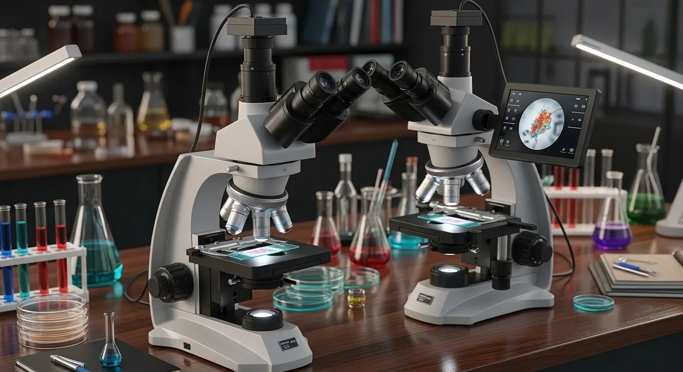
Specimen Viewing Microscopes
High Precision in the Contemporary Biological and Clinical Discovery
The role of the Specimen Viewing Microscopes in the modern scientific and medical world is central. These instruments reveal a secret world, which cannot be observed by the naked eye, from the beginning of biological specimen observation until the clinical laboratory diagnostics. It can be a tissue sample study, observation of cellular structures, or study of pathological tissues, but in whatever way, modern microscopes have revolutionized the way researchers and other medical professionals perceive life under the microscope.
These systems are not merely optical tools in the laboratories, hospitals, and research centers. They are sophisticated laboratory microscopes that are precise, keen, and deeply analytical.
What are Specimen Viewing Microscopes and Why Do They Matter?
A microscope is a scientific tool that is employed to magnify biological or medical specimens and study them using a microscope. These include:
- Biological specimen microscope.
- Clinical specimen viewing by microscope.
- Microscope of the specimen with high magnification.
These systems are significant in the analysis of microscopic specimens and examination of specimen slides that assist scientists in examining the structures that cannot be seen with the naked eye.
They are commonly used in modern laboratories in:
- Diagnosis of disease through a microscope.
- Research-based specimen visualization
- Histology slide examination
- Cytology specimen viewing
This renders them a support of clinical microscopy equipment and diagnostic microscopy equipment.
The Use of Specimen Viewing Microscopes in Labs
The knowledge of the working principle of the microscopes can be used to identify the scientific significance of the working principle of specimen viewing microscopes in laboratories. These tools are optical lenses together with sources of light and digital sensors to enlarge samples.
Key working principles:
- Light is directed through or reflected on the specimen.
- The optical microscopy technology involves the use of lenses to enlarge the image.
- Eye sharpening is a sharpening process that makes small objects sharper.
- High-resolution systems capture structural details.
Other advanced models use:
- Digital microscopy systems for real-time imaging.
- Cellular tagging by fluorescence microscopy.
- Clarity of sample phase contrast microscopy.
- Electron microscopy images of ultra-fine resolution.
It is a mixture that enables scientists to see high-resolution microscopically with the utmost accuracy.
Kinds of Microscopes for Specimen Viewing in Modern Science
The type of microscope technology needed in various labs varies according to the purpose.
Compound Light Microscopes
They are common microscopes of compounds that offer easy magnification to biological specimens.
Fluorescence Microscopes
They are used in fluorescence microscopy techniques where they are used to detect some cell structures using fluorescent markers.
Phase Contrast Microscopes
Best on clear samples, where cell structure is visible without staining.
Electron Microscopes
They provide extremely high magnifications for electron microscopy images needed in advanced research.
Practical Usage and Lab Usage
The Specimen Viewing Microscopes are needed in both clinical and research settings.
Laboratory Applications:
- Procedure of laboratory specimen analysis.
- Biological samples: Microscopic analysis.
- Methods of laboratory specimen preparation.
- Experimental microscopy procedures
Medical Applications:
- Microscopic medical diagnostics.
- Biopsy sample examination
- Cytology and histopathology.
- Cellular-level detection of diseases.
Research Applications:
- Life sciences Specimen analysis.
- Scientific microscopy applications
- Lab-based specimen imaging
- Research-grade microscope systems
These instruments offer the greatest diagnostic pathology, microscopy, and clinical specimen examination.
Modern Specimen Viewing Microscopes: Enhanced Features
The ancient microscopes were incomparable to the new ones.
Important enhanced capabilities are:
- Large specimen magnified.
- Improved microscopic magnification and resolution.
- Digital imaging integration
- AI-assisted specimen analysis
- Slide scanning systems.
- Three-dimensional microscopic imaging.
The inventions allow high-resolution microscopic analysis and accelerated diagnostic procedures in contemporary labs.
Microscopy Systems: Biological and Clinical Significance
Microscopes are significant in the study of life sciences because they can be used to study specimens.
They support:
- Learn to observe biological specimens.
- Clinical specimen diagnostic tools.
- Visualization systems of tissue specimen pathology.
- Disease diagnosis by use of histology slides.
They play a very crucial role in hospitals in detecting infections, cancer cells, and abnormalities in tissues. They are used in research laboratories to discover cell structures and biological processes.
The Significance of High Magnification in Viewing Specimens
A zoom is not merely a high magnification. It is alarming precision, conciseness, and specificity.
Benefits include:
• Improved visualization of cell structures.
• Proper analysis of tissue sample.
• Improved diagnostic confidence
• Enhanced research precision
A high-magnification microscope makes sure that the slightest biological changes are identified at an early stage.
Digital Revolution in Microscopes for Looking at Specimens
The change of the conventional optical systems to digital systems has transformed microscopy.
Digital advancements include:
• Live screens.
• Cloud-based specimen storage
• Remote laboratory access
• AI-based image analysis
These inventions have enabled the laboratory microscopy methods to be more efficient, quicker, and accessible worldwide.
TrustLab-China and Advanced Biological Microscopes
In modern laboratories, quality equipment is normally used to ensure that the appropriate results are obtained. TrustLab China is one of the reputable suppliers of advanced laboratory equipment with a reputation for precision-engineered solutions. These systems are directed at:
• Clinical laboratory diagnostics
• Research-based specimen visualization
• Proper observation of biological samples.
• State-of-the-art imaging of lab samples.
They may be applied in scientific microscopy as well as in clinical laboratory diagnostics, thus they can be applied in hospitals, universities, and research institutes.
Future of Specimen Viewing Microscopes
Technology is the future of microscopy. Expected developments include:
- Artificial intelligence diagnostic microscopy systems.
- Improved 3D tissue imaging.
- Laboratory intelligent microscopy.
- Robotic specimen-analysis devices.
The innovations will also enhance the visualization of medical specimens and speed up the process of detecting the disease.
Conclusion
Specimen viewing microscopes are not just a laboratory tool, but a window into a world that is not visible. They allow scientists and doctors to observe what was not visible before because of the examination of biological specimens, up to high-tech clinical diagnostics.
The changing technology, such as digital imaging, fluorescence systems, and AI-assisted analysis, is also giving microscopy a new spin towards accuracy and clarity.
The quality specimen viewing microscopes produced by TrustLab China, in case you are interested in modernizing your laboratory with efficient and quality solutions, are precise, innovative, and scientifically superior. Determine their solutions today and add microscopic clarity to your research world.