


Updated 1 month ago
How Dual LED Lighting Works in a Laboratory Microscope with LED Illumination
Arafatshuvo
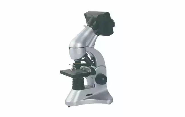
Laboratory Microscope with LED Illumination
For a Laboratory Microscope with LED Illumination, many factors besides magnification impact image quality. These factors include the direction of illumination, the level of transparency of the specimen, the performance of the objective, the exposure settings of the camera, and the system that displays the image.
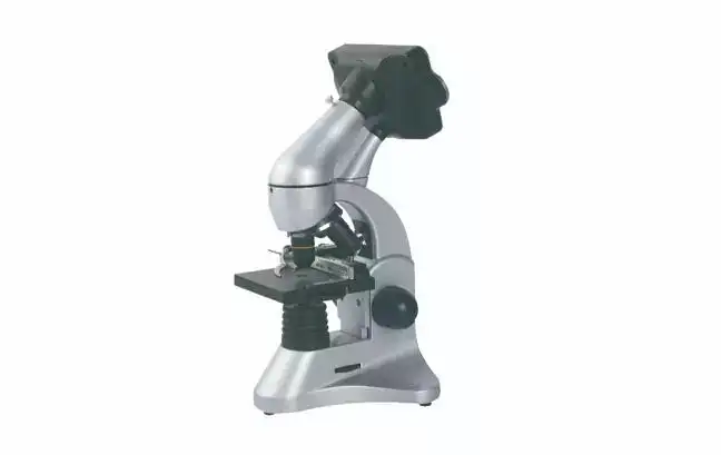
At Trustlab, we treat illumination as part of the complete optical system rather than as a standalone light source. This is particularly important in a digital biological microscope using both upper and lower LEDs.
The Trustlab GT-GXSP-45LCD Digital Biological Microscope combines 40X–400X optical magnification, dual LED illumination, a 2.0 MP CMOS camera, built-in LCD display, SD storage, and USB connectivity. Its configuration illustrates how a Laboratory Microscope with LED Illumination can support both direct observation and digital documentation.
Why Dual LED Illumination Matters
The first question is not "How bright is the LED?" but:
Where should the light come from for this specimen?
A dual-LED microscope uses two illumination paths:
| Illumination Mode | Light Direction | Best Starting Point For | Main Purpose |
| Lower LED | Through the specimen | Slides, cells, thin tissue sections | Transmitted-light observation |
| Upper LED | Onto the specimen surface | Opaque or surface-dominant specimens | Reflected-light observation |
| Dual-light configuration | Selectable upper/lower paths | Mixed teaching and laboratory samples | Wider specimen compatibility |
The lower LED produces transmitted illumination. Light passes through the specimen before entering the objective, which is typically required for transparent or semi-transparent biological samples.
The upper LED works differently. Light is directed onto the sample surface and reflected back into the optical system. This approach is useful when the specimen does not transmit sufficient light.
Therefore, the advantage of a Laboratory Microscope with LED Illumination using two light paths is not simply higher brightness. It is the ability to match illumination geometry to specimen structure.
Why Higher Magnification Often Requires More Careful Lighting
A frequent operating problem occurs when a specimen looks bright at low magnification but becomes darker or loses contrast at higher magnification.
This does not necessarily indicate insufficient microscope power.
Moving from lower magnification toward 400X changes several conditions simultaneously:
• The field of view becomes smaller;
• Focusing becomes more sensitive;
• Optical alignment becomes more critical;
• Available image brightness may decrease;
• Camera exposure becomes more sensitive to LED intensity.
The correct response is not always to increase LED output.
In microscopy, brightness and resolution are different parameters. Resolution depends strongly on the objective optics and numerical aperture, while illumination determines whether available detail can be observed with sufficient contrast.
For this reason, a professional Laboratory Microscope with LED Illumination should be evaluated as an optical system rather than by LED brightness or magnification alone.
LED Intensity, Contrast and CMOS Exposure
Digital microscopes introduce another variable: the camera sensor.
The GT-GXSP-45LCD uses a 2.0 MP CMOS sensor with 1600 × 1200 image resolution. Light reaching the CMOS sensor must be sufficient for imaging, but excessive illumination can produce clipped highlights and washed-out fine structures.
The imaging chain can be understood as:
LED illumination → Specimen → Objective → Optical image → CMOS sensor → LCD → Image storage
Increasing LED intensity affects the sensor exposure, but it does not increase optical resolution.
This distinction becomes important when operators use the system's 8X digital zoom. Digital zoom enlarges recorded pixels; it does not provide the same information gain as increasing optical resolution.
For procurement teams comparing a Laboratory Microscope with LED Illumination, three specifications should therefore be evaluated separately:
• Optical magnification;
• Camera resolution;
• Display resolution.
They should never be combined into one "effective magnification" figure.
How the GT-GXSP-45LCD Integrates Optics and Digital Imaging
Trustlab designed the GT-GXSP-45LCD as a compact digital biological microscope for routine laboratory, education, and observation workflows.
Its main specifications include:
| Parameter | GT-GXSP-45LCD Specification |
| Optical magnification | 40X–400X |
| Eyepiece | WF10X |
| Objectives | Achromatic, up to 40X |
| Illumination | Upper and lower LED |
| Camera | 2.0 MP CMOS |
| Image resolution | 1600 × 1200 |
| LCD | 3.6-inch TFT |
| LCD resolution | 960 × 240 |
| Digital zoom | 8X |
| Internal memory | 32 MB |
| External storage | SD card |
| Interface | USB 2.0 Mini |
| Power | DC 5V / 1A |
These values also demonstrate why microscope specifications must be interpreted correctly.
For example, the 1600 × 1200 camera resolution and 960 × 240 LCD resolution describe different parts of the system. The camera determines captured pixel dimensions, while the LCD determines how the image is displayed locally.
The same principle applies to optical and digital magnification.
Dual LED vs. Single LED Illumination
A single lower LED is often sufficient for laboratories that work almost exclusively with prepared transparent slides.
Dual illumination becomes more relevant where sample types vary.
A dual-LED system can be useful when users need to:
• Observe both transmissive and surface-dominant samples;
• Switch between biological slides and thicker teaching specimens;
• Reduce dependence on a single illumination geometry;
• Perform direct observation and digital image capture on the same platform.
However, dual illumination should not be marketed as automatically producing higher resolution. Its main technical value is specimen compatibility and illumination flexibility.
LED vs. Halogen: Practical Laboratory Differences
LED illumination has become common in laboratory microscopy because it can simplify routine operation.
Compared with traditional halogen systems, LED illumination can offer:
• Lower heat generation around the microscope stage;
• Reduced lamp replacement requirements;
• Lower electrical consumption;
• Faster start-up;
• More convenient routine brightness control.
Halogen sources can still be appropriate in specific optical systems, so the correct choice depends on application requirements rather than a simple "LED is always better" rule.
Setup and Maintenance of a Laboratory Microscope with LED Illumination
For consistent results, Trustlab recommends treating illumination setup as part of the observation procedure.
A practical workflow is:
- Identify whether the specimen is transparent, semi-transparent, or opaque.
- Select the upper or lower LED accordingly.
- Start at lower magnification.
- Bring the specimen into focus.
- Increase magnification as required.
- Adjust LED intensity only after the optical image is properly focused.
- Check the CMOS image for overexposure before recording.
Optical surfaces should remain clean, and internal LED, camera, or electronic modules should not be disassembled during routine maintenance.
For technical procurement, buyers should also confirm parameters not always shown in a basic product specification, such as LED color temperature, rated lifetime, objective numerical aperture, illumination adjustment range, and available compliance documentation.
Choosing the Right Laboratory Microscope with LED Illumination
A sound microscope selection process should follow this sequence:
Specimen → Illumination → Optics → Digital imaging → Workflow → Maintenance → Compliance
For laboratories, schools, distributors, and equipment projects requiring flexible illumination and integrated digital observation, the Trustlab GT-GXSP-45LCD provides a practical combination of 40X–400X optical magnification, upper and lower LED illumination, 2.0 MP CMOS imaging, LCD viewing, SD storage, and USB connectivity.
When these specifications match your sample types and workflow, the next step is to review the complete configuration and technical documentation for the GT-GXSP-45LCD directly with Trustlab.
FAQs
Q1. What is the magnification range of Trustlab's Laboratory Microscope with LED Illumination?
The Trustlab GT-GXSP-45LCD provides a 40X–400X magnification range, using a WF10X eyepiece and achromatic objectives up to 40X. This range is designed for routine biological observation, education, training and general laboratory work.
Q2. Why does the Trustlab microscope use both top and bottom LED illumination?
The top and bottom LED illumination allows users to adapt the lighting direction to different specimen characteristics. Bottom illumination is generally used when light needs to pass through a specimen, while top illumination can support observation of surface-oriented or less transparent samples.
Q3. Does the Trustlab GT-GXSP-45LCD require a computer for observation?
No. The microscope integrates a 3.6-inch TFT LCD, allowing real-time observation directly on the instrument without requiring a continuously connected external computer.
Q4. What camera does the Trustlab digital biological microscope use?
The GT-GXSP-45LCD uses an integrated 2.0 MP CMOS camera for digital image capture. Trustlab specifies an image resolution of up to 1600 × 1200 pixels.
Q5. Is the 8X digital zoom the same as additional optical magnification?
No. The microscope provides 8X digital zoom, but digital zoom enlarges the captured image rather than increasing the resolving capability of the microscope optics. Trustlab distinguishes digital enlargement from the system's 40X–400X optical magnification.