


Updated 1 month ago
Microscopes for Laboratory Testing: Binocular vs. Trinocular Designs for Modern Labs
Arafatshuvo
Microscopes for Laboratory Testing
When specifying Microscopes for Laboratory Testing, start with the specimen and the required output: visual assessment, a reference photograph, a calibrated measurement or a live teaching display. Each places different demands on the optical and imaging system.
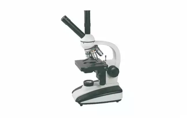
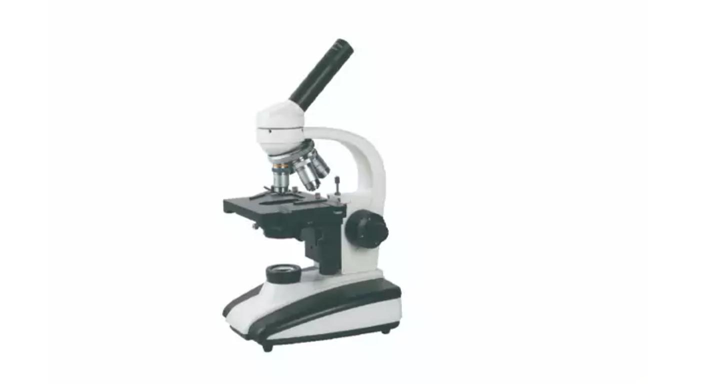
At Trustlab, our GT-XSP-136B provides a binocular configuration for routine observation. To compare it meaningfully with a trinocular system, buyers should examine the complete workflow—including camera access, optical performance, maintenance and acceptance testing.
1. What is the Difference: Binocular vs. Trinocular?
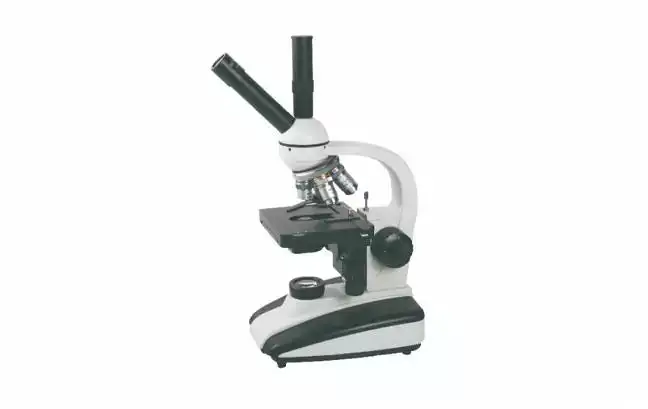
Binocular and trinocular head designs are similar in that they both incorporate two eyepieces to facilitate binocular viewing. The difference lies in a trinocular head design which incorporates an additional photographic path, using a prism or beam splitter to redirect a portion of the light to a photographic instrument.
A third port does not increase objective resolution. Its value is camera access.
Depending on the head design, light may be switched between the eyepieces and camera or divided between them. Therefore, "trinocular" alone does not confirm simultaneous viewing and photography. Request the available light-distribution modes and confirm which output each stated ratio describes.
A compatible eyepiece camera may allow photography through a binocular microscope, but it can occupy one viewing position. Mechanical fit, optical coverage and operating convenience require verification.
2. Select Microscopes for Laboratory Testing Based on Your Workflow
Most Microscopes can be specified using the following parameters.
| Parameter | Binocular Microscopes | Trinocular Microscopes | Notes |
| Routine inspections | Direct view of specimen | Direct view of specimen | Compare hand-feel of optics |
| Occasional Photographs | Eyepiece camera may be sufficient | Dedicated photography port | Estimate disruption, setup time |
| Routine Documentation | Camera may obstruct specimen view | Dedicated port may be better for repeated views | Estimate in-focus and correct exposure |
| Teaching | May obstruct view for specimen adjustment | May be used for simultaneous display | Demonstrate specimen in actual view |
| Dim specimens | May require adjust exposure | May require beam splitter | Choose path for better contrast |
| Ownership Costs | Reduced optomechanical complexity | Add'l cost for adaptors, camera, and software | Assess total costs |
Imaging Capability or Frequency? With Microscopes for the Lab, it's important to consider both. For example, the Lab may need to take a lot of reference photos or perform documentation, justification for a more expensive purchase.
3. Evaluate Resolution, Illumination and Mechanics
Magnification Is Only One Specification
Visual magnification equals eyepiece magnification multiplied by objective magnification. A 10X eyepiece and 40X objective provide 400X.
Resolution also depends on objective numerical aperture, optical correction, specimen preparation and illumination. Increasing displayed image size cannot reveal detail the optical system has not resolved.
For camera documentation, evaluate field flatness as well as central sharpness. An image that looks acceptable at the centre may show edge defocus across a larger sensor.
Trustlab's Binocular Configuration
Our published GT-XSP-136B specification provides the following reference for comparing Microscopes for Laboratory Testing:
| Component | Specification | Selection relevance |
| Eyepieces and objectives | WF10X; achromatic 4X, 10X, 40Xs, 100Xs oil | 40X–1000X visual magnification |
| Viewing head | Binocular, 45° inclined, 360° rotating | Assess posture and positioning during trials |
| Illumination | 6V/20W halogen | Include replacement lamps in maintenance planning |
| Condenser | NA 1.25 Abbe, iris diaphragm and filter | Provides illumination-aperture adjustment |
| Stage | 115 × 125 mm, double-layer mechanical | Evaluate slide handling and specimen positioning |
| Focus | Coarse and fine adjustment; 20 mm range | Verify smooth adjustment and focus stability |
These are configurations, rather than measured acceptance results. Condenser NA is not objective NA; stage dimensions are not travel distances; focus range is not fine-focus resolution.
4. Consider the Entire Optical Path When Choosing a Camera
Consider the following when thinking about cameras other than connectivity.
•Image Scaling: There has to be an adapter for each camera to show images at the right scale.
•Image Sensor Coverage: The camera has to be able to capture the entire image field of view.
•Sampling: The scale factor of image has to be determined to set the physical size of the image sensor and pitch of the images sensor elements.
•Parfocality: To be useful, the focal planes of the camera and eyepiece must be coplanar.
•Software: The operating system and the camera must be compatible and allow control of the camera functions. Images must be captured in formats of usefulness and measurements must be taken in appropriate units.
In the absence of other constraints, greater image sensor size results in larger captured field of view. Camera field of view can be less than the eyepiece field of view. Smaller projection magnifications capture larger fields of view and greater magnifications capture smaller fields.
Request images from the proposed objective–adapter–camera combination, including both the centre and edges.
5. Establish Installation and Acceptance Checks
Microscopes will be inspected against defined criteria to determine acceptability prior to laboratory testing.
•Installation Checks: checks include, but are not limited to, verification of support, attachment, and electrical supply.
•Observation Checks: checks to be performed include, but are not limited to, adjustment of controls and evaluation of illumination and image quality.
•Mechanical Checks: evaluation of stage and focus controls.
•Imaging Checks: Evaluate image quality, exposure and file export.
Calibrate image scale to evaluate and verify measurement. Revalidate image scale if change is made to the adapters, optics or other components that affect image size. Retain all relevant information pertaining to the acquisition of the image.
Additional checks may include, but are not limited to, cleaning the microscopic objectives using the manufacturer's recommended procedure, and dust covering objectives, in accordance with laboratory procedures.
6. Specify a Defined Configuration
Microscopes will be compared using an itemized quote for the system (microscope, adapters, camera, and related software and accessories), calibration, consumables, and support. Requests for documentation will be made for the microscopes, and related evidence of conformity and warranty.
We list OEM availability for the GT-XSP-136B; customization scope requires project confirmation. The published configuration does not establish trinocular conversion or camera compatibility.
Explore Trustlab's laboratory microscope and send us your specimen types, observation requirements and recording frequency. These details provide a practical basis for confirming the model and accessory scope before purchase.
FAQs
Q1. Is Trustlab's GT-XSP-136B a binocular or trinocular microscope?
The GT-XSP-136B is a binocular microscope with a 45° inclined, 360° rotating viewing head. A trinocular configuration is not listed on its product page.
Q2. What magnification does Trustlab's GT-XSP-136B provide?
It provides 40X–1000X total visual magnification using WF10X eyepieces and 4X, 10X, 40Xs and 100Xs oil-immersion objectives.
Q3. Which laboratory applications does Trustlab list for this model?
Listed applications include routine specimen observation, biological slide examination, microscopy training and basic research. Confirm suitability for your specific testing method before purchase.
Q4. Does the Trustlab microscope include an oil-immersion objective?
Yes. The listed configuration includes a 100Xs oil-immersion objective. Follow the operating instructions for compatible immersion oil and lens cleaning.
Q5. What illumination system does Trustlab use in this model?
The GT-XSP-136B uses a 6V/20W halogen lamp and an NA 1.25 Abbe condenser with an iris diaphragm and filter.