Updated 2 months ago
Tissue Specimen Observation in Histology | Methods & Laboratory Guide
sidrahafeez754

Tissue Specimen Observation
Understanding Tissue Specimen Observation in Biological and Medical Studies
One of the most crucial procedures in the medical sciences, histology, and pathology diagnostics is Tissue Specimen Observation. It is concerned with the study of biological tissues on both microscopic and structural levels in order to determine normal and abnormal cellular behavior.
In a contemporary laboratory, laboratory observation of tissues is not only a matter of looking at slides. It is a complete scientific procedure, which entails preparation, fixation, sectioning, staining, and finally, extensive microscopic examination. In its simplest form, observation of histological tissue specimens aids scientists in comprehending the behavior of cells in health and disease.
Step-By-Step Tissue Specimen Observation Process in Histology
The step-by-step observation of the tissue specimen is highly systematic and follows the laboratory procedures. All these steps ensure that the biological structure is not ruined so that it can be analyzed properly.
1. Tissue Fixation Process
The initial level is preservation. Formaldehyde and other chemical fixatives are applied to prevent decay in tissues and maintain the structure of cells.
This is a significant measure to:
• Preventing tissue degradation
• Maintaining protein structure
• Helping in the histological study of tissue samples.
2. Paraffin Embedding Method
Embedding of tissues in paraffin wax is followed by fixation. This enables thin slicing without destroying the structure.
This step aids in the observation of paraffin-embedded tissue sections that are popular in diagnostic pathology.
3. Microtome Sectioning
The embedded tissue is cut into ultra-thin slices using a microtome. These are put on glass slides to be examined.
4. Slide Preparation Techniques
Stained ready parts are mounted. The methods of adequate slide preparation are required to give clarity in the microscopic analysis of tissues.
5. Staining Protocols (H&E Staining & Special Stains)
Staining is one of the most significant steps:
- Hematoxylin: Staining of nuclei is blue.
- Eosin is pink in the cytoplasm.
- It is known as hematoxylin and eosin staining observation, which is a gold standard in histopathology.
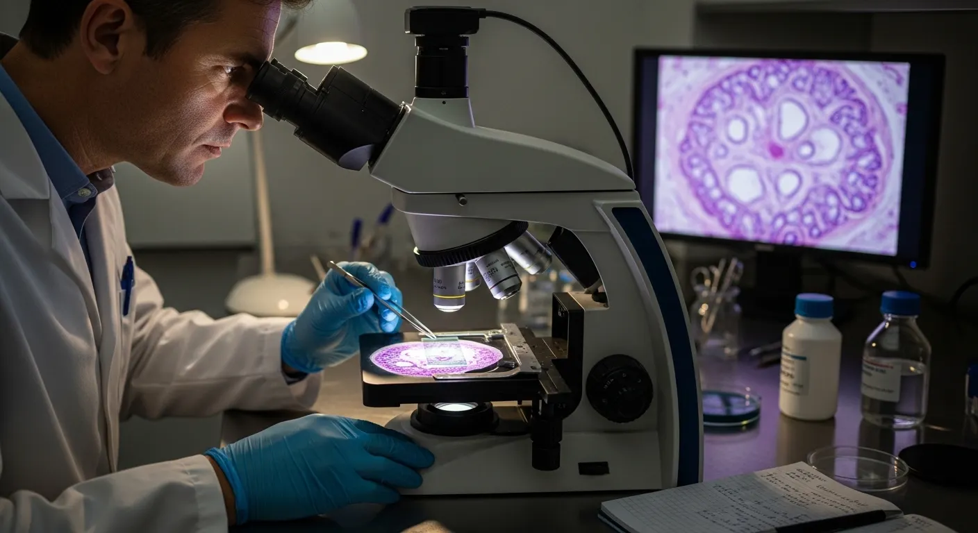
Cellular Morphology and Histological Examination
After the preparation of slides, specialists start to examine them under a microscope.
This includes:
• Cellular morphology assessment
• Tissue section evaluation
• Structural tissue analysis
• Biological tissue examination
At this point, scientists are interested in:
• Cell shape and size.
• Nucleus integrity
• Tissue organization
• Abnormal growth patterns
Microscopic tissue examination is geared towards identifying the tiniest changes in the tissue structure.
Microscopic Methods of Observation of the Tissue Specimen
In modern laboratories, microscopic observation of tissue specimens is performed with advanced imaging techniques.
Light Microscopy Observation
This is the most common way of routine tissue examination.
Electron Microscopy Imaging
Applications: Cellular structure imaging with ultra-high resolution.
Digital Pathology Imaging
Remote viewing and analysis of slides is possible with digital systems.
These techniques enhance:
• Magnified cellular structure.
• Microscopic slide visualization
• High-resolution tissue imaging
• Cellular architecture examination
Tissue Sample Analysis and Diagnostic Pathology Workflow
Microscopic analysis of tissue samples is important in diagnosis in the clinical setting.
The workflow of pathology diagnosis consists of:
• Biopsy collection
• Laboratory specimen processing
• Slide preparation
• Histopathological observation
• Diagnostic tissue review
This workflow helps in:
• Disease tissue identification
• Abnormal cell detection
• Tumor tissue analysis
• Study of inflammatory tissue response.
• Study of necrosis and degeneration.
Clinical Significance of Tissue Specimen Examination
The clinical tissue specimen observation process is crucial in the healthcare system.
It supports:
• Early cancer detection
• Chronic disease monitoring
• Infection identification
• Treatment planning
The interpretation of a patient's biopsy is used by the physicians to make life-saving decisions.
Key benefits include:
• Faster diagnosis
• Accurate disease classification
• Improved patient outcomes
This is the reason why biomedical tissue research is ever-increasing around the world.
Histology Equipment and Laboratory
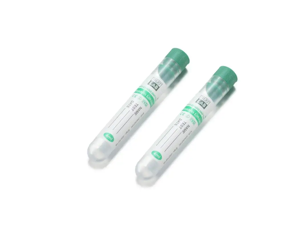
To guarantee accurate results in the examination of tissue specimens, proper laboratory equipment is required. A histology cassette is one of the tools that are important in processing samples. Histology cassettes are used to:
• Safe tissue storage
• Organized processing
• Fixation chemical resistance.
• Efficient laboratory workflow
Other tools that are required are:
• Microtomes
• Embedding stations
• Staining jars
• Slide racks
These instruments enhance the methods of observation of tissues in the laboratory and guarantee accuracy.
Background in Tissue Viewing, Histology, and Pathology
Tissue Specimen Observation is closely related to histology and pathology.
It involves:
• Histology laboratory procedures
• Pathology tissue analysis
• Disease tissue identification
• Tumor evaluation
• Inflammatory response study
Histopathological observation helps experts to comprehend the way the disease progresses at a cellular level.
This helps in:
• Early diagnosis
• Research development
• Drug testing
• Clinical advancements
Biomedical and Scientific Importance of Tissue Observation
Biomedical science uses tissue observation to:
- Biomedical tissue research
- Laboratory diagnostics in histopathology.
- Identification of disease markers in tissues.
- Medical specimen investigation
It gives us an insight into:
- Cellular tissue organization
- Tissue structure analysis
- Structural abnormalities
- Disease progression patterns
This renders the study of biological tissues a pillar of contemporary medicine.
Tissue Samples Interpreted by Microscopy
Microscopy is an important aspect of tissue morphology in microscopes. It helps researchers to perform:
• Microscopic slide visualization
• Cellular architecture examination
• High-resolution tissue imaging
• Structural comparison studies
By such techniques, researchers carry out cellular morphology analysis with accuracy.
State-of-the-Art Laboratory Workflow in Histopathology
The complete laboratory process involves:
• Sample collection
• Tissue fixation process
• Embedding and sectioning
• Staining protocols
• Microscopic analysis
• Diagnostic interpretation
It is a methodical procedure that offers quality histological analysis of the tissue samples.
Importance of Tissue Specimen Observation in Histology
This process cannot be overestimated.
It is used for:
• Clinical diagnosis
• Medical research
• Pharmaceutical development
• Disease prevention strategies
In its absence, there would be no modern pathology.
Conclusion:
Tissue specimen observation is not only a laboratory process, but the basis of modern diagnostic science. Ever since the microscopic analysis of tissues to histopathological analysis, every step reveals some hidden truths about human health.
The field is evolving fast with sophisticated tools, sophisticated staining procedures, and accurate lab processes. The contemporary laboratories rely on adequate analysis of the tissue section to make a life-saving diagnosis.
Trustlab China has the answer to the contemporary labs in case you are interested in modernizing your histology workflow.